Head Case

Update, Dec. 4, 2014: In the last week the human brain collection has received a flurry of regional and national media attention, and on Wednesday UT released a statement saying that the brains missing from the collection had been thrown out. "University officials will appoint a broader investigative committee to examine these issues," the statement said. Below is our original story from last year, which focused more on the research applications than the whodunnit.
For more than 20 years, a collection of nearly 100 human brains went unnoticed, lining the shelves of a closet somewhere in the back of a UT animal lab. Uncharacteristically small, large, or disfigured, these brains were all considered abnormal.
The unique specimens were preserved from patients at the Austin State Hospital, some dating back to the 1950s. The only remaining records were brief descriptions on the jar labels like “Down's syndrome" and “hydrocephalus,” indicating water on the brain. In the midst of relocating and storing the brains, their original files have been lost.
In 2011, photographer Adam Voorhes came to the University to borrow a brain from the psychology lab of professor Tim Schallert for an upcoming article in Scientific American. Before departing, Schallert asked him if he wanted to see one more thing.
As he was led to the the cerebral gold mine, Voorhes was confused. Why had the brains been gathering dust for so many years at an institution rife with capable researchers right outside the door?
In 1985, the hospital had realized it was in violation of federal guidelines regarding preservation methods and was forced to find a new home for the brains. UT eventually acquired them after a fierce battle with other institutions, including Harvard. However, UT lacked the proper machines or technology to handle the brains and due to a lack of funding, they sat forgotten for more than two decades.
"If you've got an intact brain, you have a conundrum. You don't want to destroy it if you don't know what you are looking for," says Larry Cormack, a UT psychology professor and curator of the brains. "Once we got a high-tech scanner on campus, it made a difference. Now we have the technology to work with them."
The new high-resolution MRI scanner was delivered to campus in February 2012 and was put in use by May.

Along with the scanner's arrival, Voorhes' plan to photograph the entire collection helped spur the University to finally make use of the valuable collection.
The brains were handed over to the Freshman Research Initiative, an undergraduate program where students have the opportunity to gain hands-on lab experience. After completing a lab basics course, students in the brain-pathology sequence will get to handle the brains directly.
"The freshmen are really excited to help scan," Cormack says. "A lot of them are pre-med and now they are on the cutting edge to take a sneak peek into the brains."
This spring, students completed MRI scans for two of the brains and hope to complete the rest this fall.
What makes these preserved brains special is their ability to be scanned multiple times and for long hours— something that a living brain wouldn't be able to withstand.
"This is a remarkably large collection of intact brains, and we don't know which one is the most exciting," Cormack says. "That is the reason for these scans—to finally see inside."
The only trouble the research team faces is the lack of documentation associated with each brain.
"Since we don't have their medical records, even if we find anything, it potentially could be useless if we do not have a human match," says Mithra Sathishkumar, a teaching assistant and research educator for the Freshman Research Initiative.
The group hopes to compile a database of information for laboratory use and share it with other institutions conducting similar work. "Once word gets out that we have these brains, people will be interested," Cormack says.
Upon conclusion of the study, some of the brains will be put on permanent display in the Image Researching Center on campus.
The full collection of Adam Voorhes' photos can be found here.
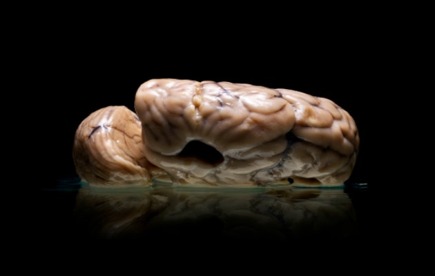
Top: Brain Study no. 331, Severe Developmental Anomaly of the Brain, 1978.
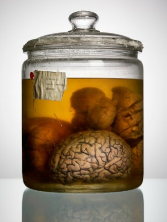
Bottom: Brain Study no. 9, Down's Syndrome, 1983. Both photos courtesy Adam Voorhes.


